

Summary
- Nikon has announced the LS-X for three-dimensional imaging of living biological specimens.
- It works with the ECLIPSE Ti2-E microscope using light sheet illumination.
- The system supports time-lapse imaging, organoids and complementary microscopy methods.
- Phased availability begins on November 30, 2026.
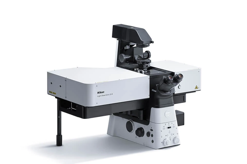
Nikon has introduced the LS-X Light Sheet Unit, a system that builds detailed three-dimensional images of living biological samples from successive microscopic optical sections.
Designed for the ECLIPSE Ti2-E inverted research microscope, it lets scientists examine the interiors of organoids, embryos and other three-dimensional structures, including as they change over time. Nikon announced the LS-X on November 12, 2026, with phased availability planned from November 30.
How a thin sheet of light creates a 3D image
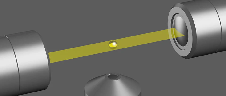
In light sheet microscopy, a thin plane of illumination selectively excites the section of the specimen being captured by a camera. Images collected at successive depths can then be assembled into a three-dimensional representation of the specimen’s interior.
The LS-X illuminates samples from two sides to help reveal structures within thicker three-dimensional specimens. Restricting excitation near the focal plane reduces out-of-focus fluorescence background and limits unnecessary light exposure elsewhere in the sample.
From brain organoids to living cell cultures
Organoids are small, three-dimensional cell cultures that reproduce selected characteristics and functions of real tissues. Studying their development and activity requires a view beneath the surface while allowing observation to continue over time.
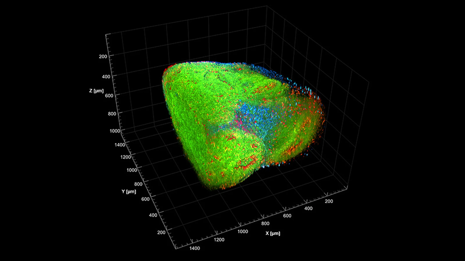
Nikon’s launch materials include a 3D image of a brain organoid, using a specimen supplied by Professor Yoshiho Ikeuchi’s laboratory at the University of Tokyo. The LS-X also supports tissue-cleared samples, in which scattering has been reduced, and expansion-microscopy specimens, physically enlarged to make finer structures easier to examine.
Recording change over time
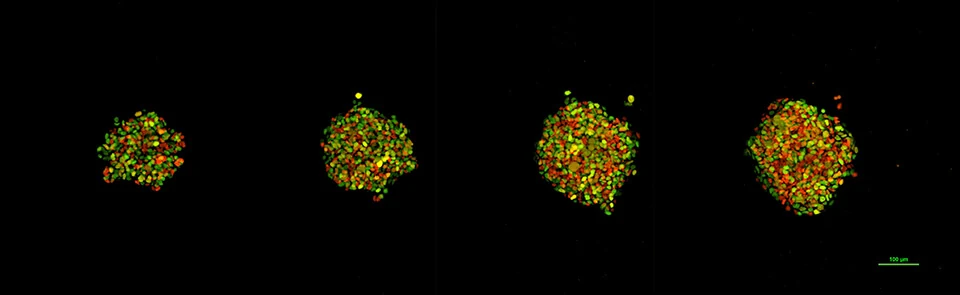
The LS-X is not limited to static 3D reconstructions. Lower unnecessary light exposure matters when living structures must be imaged repeatedly during long time-lapse experiments. The system also supports near-infrared excitation, while a dedicated Tokai Hit incubator can maintain conditions suitable for extended live imaging.
One example is a HeLa-cell-derived spheroid shown over four days, from day 0 to day 3. Its fluorescent colors come from Fucci reporters that track cell-cycle progression. These are therefore visual representations of biological signals rather than colors the unaided human eye would see in the specimen.
One microscope, several imaging methods
The ECLIPSE Ti2-E platform can support different microscopy methods, including light sheet, confocal and epifluorescence imaging. With a compatible configuration, laboratories can add LS-X capabilities to an existing instrument rather than set up a separate microscope for every technique.
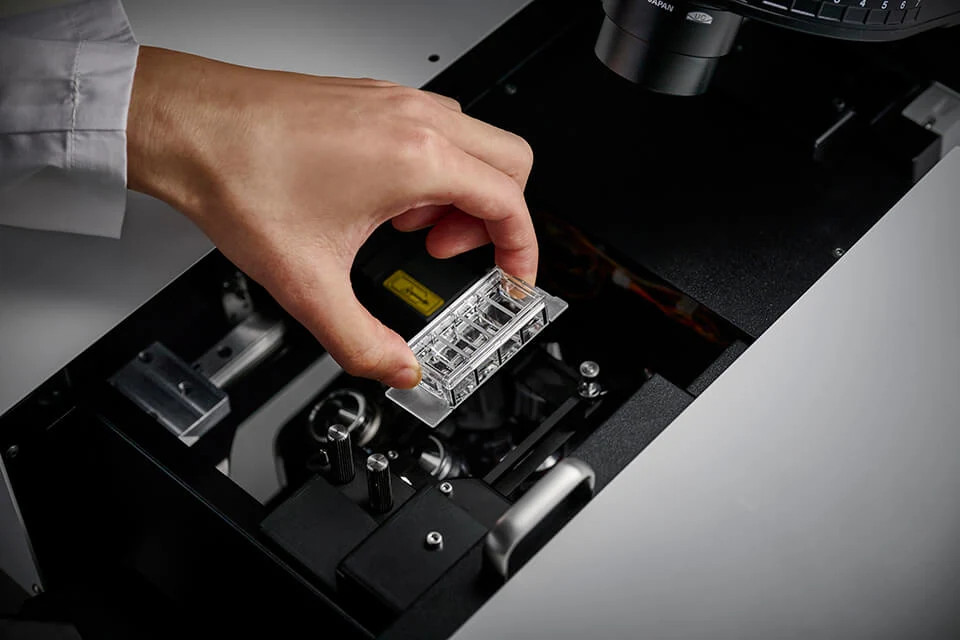
NIS-Elements software automates light sheet optical alignment, while dedicated multi-chamber slides allow culture, preparation and imaging with fewer transfers between vessels. The same sample can also be examined using complementary microscopy methods.
The connection between laboratory instruments and photographic practice is also clear in the Nikon Small World 2026 winning photographs, where fluorescence, confocal microscopy and other methods reveal subjects that a conventional camera could not capture in the same way.
Price and availability
Nikon plans to begin phased availability of the LS-X on November 30, 2026 through its regional distributors. No official European price or specific availability date for Greece was identified.
More information here.
What we think
The most striking microscopy images can look like scenes from another planet. What makes this system particularly interesting, though, is that Nikon is trying to record what happens inside a living specimen while that specimen continues to change, not merely produce an attractive 3D rendering.
For image makers, the interplay of illumination, focus and time is fascinating. Just as in photography, light determines what we can see. In microscopy, it can also affect the thing being observed. Selective excitation is therefore much more than an optical specification: it can determine whether a living experiment yields useful images over many hours.
These images also need to be read correctly. A 3D view is assembled from multiple optical sections, and vivid colors often encode signals from fluorescent markers. That does not make the pictures less remarkable. It highlights how creative and carefully documented scientific imaging can be.
Frequently asked questions
Does the Nikon LS-X take a 3D photograph in a single shot?
Not in the sense of a conventional camera exposure. Its 3D representation is assembled from images captured at successive depths within the specimen.
Can the LS-X be added to an existing Nikon ECLIPSE Ti2-E?
Nikon states that compatible ECLIPSE Ti2-E installations can add light sheet imaging with the LS-X. Exact compatibility depends on the configuration of the microscope.