

Summary
- Nikon announced the 2026 Small World winners.
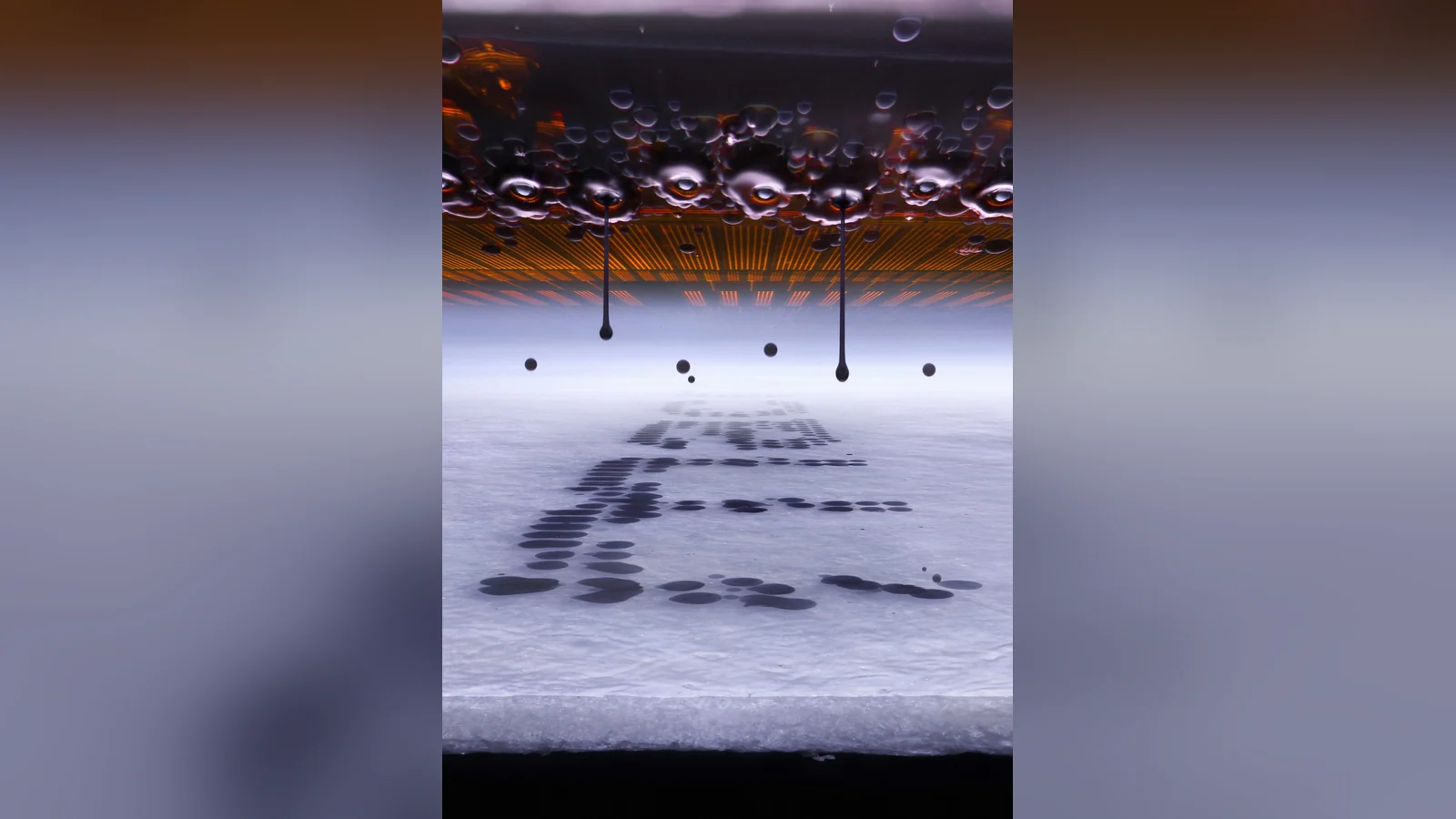
- Ole Bielfeldt took first place with ink droplets inside a printer.
- The competition received 1,803 entries from 69 countries.
- This gallery presents the top 20 and nine honorable mentions.
What happens when we look at an ink droplet, a spider or a neuron through a microscope? The Nikon Small World 2026 award-winning photographs reveal details that the naked eye cannot see.
The 52nd edition drew 1,803 submissions from 69 countries. Nikon recognized 86 photographs overall, including 20 ranked winners and additional distinctions. This gallery presents all 29 supplied photographs: the complete top 20 and nine honorable mentions.
First place: a microscope inside a printer
Cologne-based experimental photographer and macrofying creator Ole Bielfeldt photographed an inkjet printer depositing ink onto paper. To overcome the speed of the moving printhead, he modified the printer so that its head remained stationary while the paper moved. A microscope objective and flash let him freeze the ink droplets in flight.
The homemade rig used conventional techniques including image stacking and image processing, rather than generative AI image creation. The approach carries added relevance after Nikon revoked the first prize in its separate Small World in Motion 2026 video competition for an AI rules violation.
The top 20 award-winning photographs
1st place — Ole Bielfeldt. Ink droplets during inkjet printing.
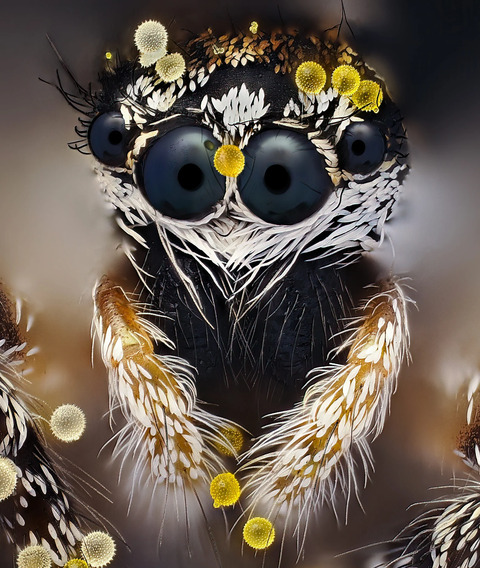
2nd place — John-Oliver Dum. Jumping spider (Salticus scenicus) with pollen.
3rd place — Kaelin Wulf και Rachel Flannery. Neurons in an expanded mouse brain.
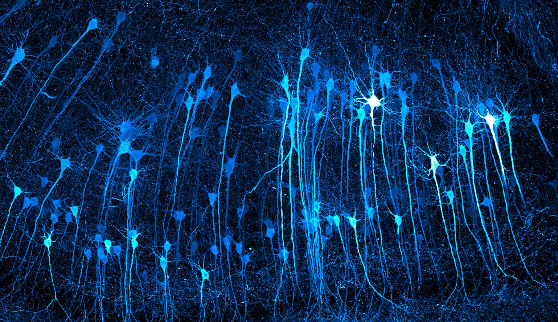
4th place — Justin Theodore Benttinen. Tip of a ballpoint pen.
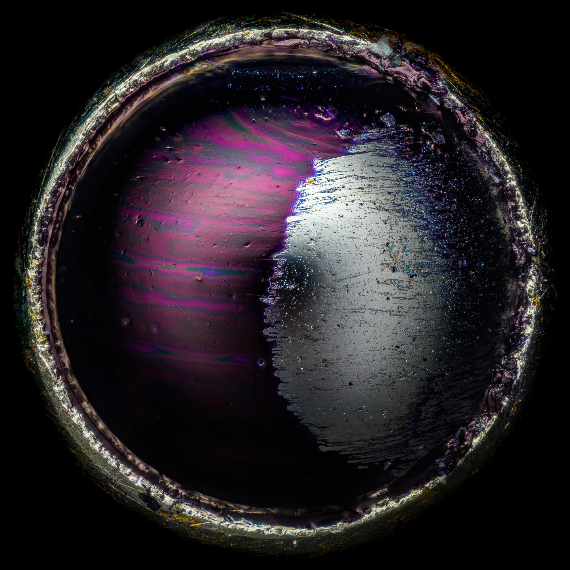
5th place — Mike Crutchley. Internal structure of a closed daisy.
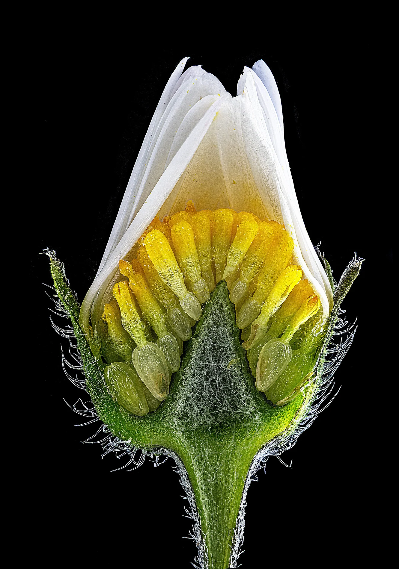
6th place — Eric Epstein. Hemitrichia slime mold fruiting bodies.
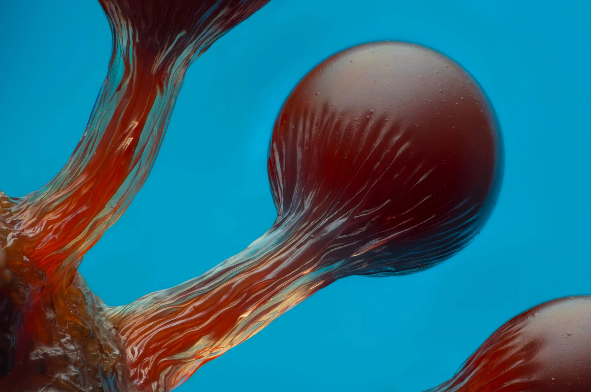
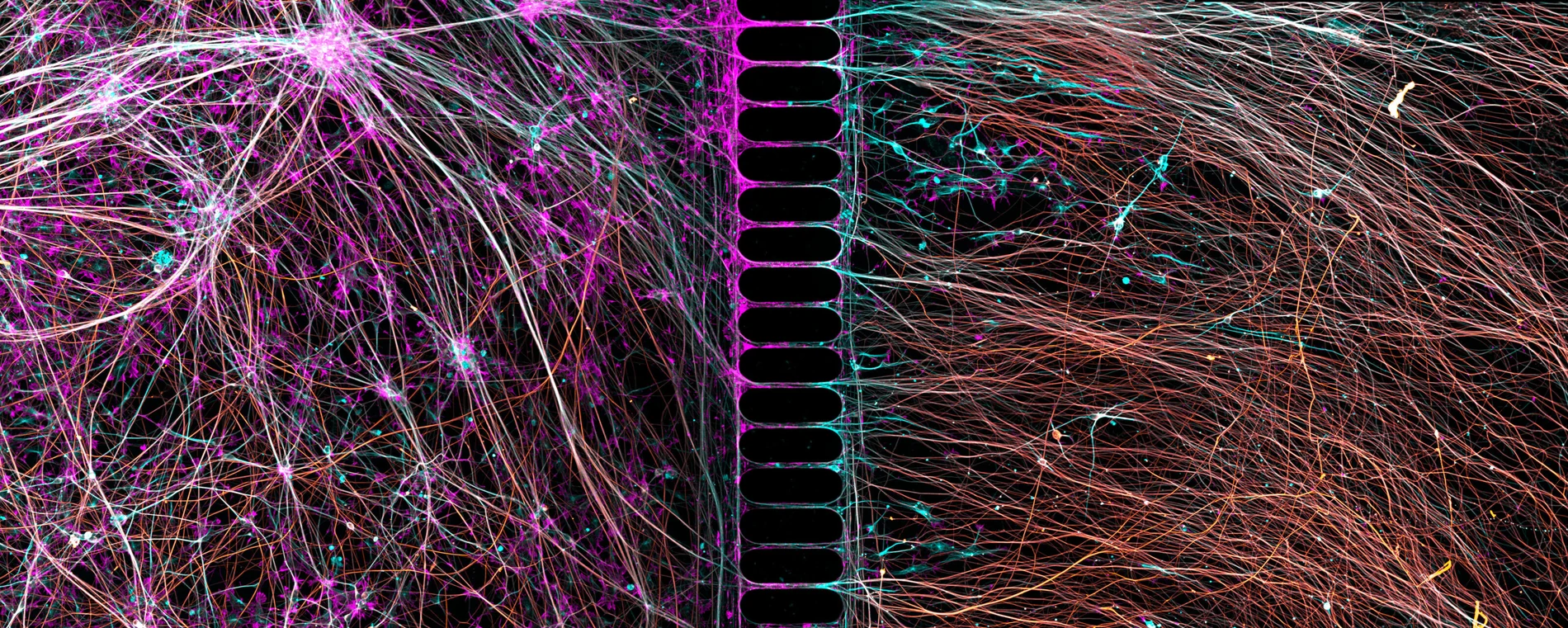
7th place — Bruno Cisterna και Eric Vitriol. Human neurons reprogrammed from skin cells in a microfluidic chamber.
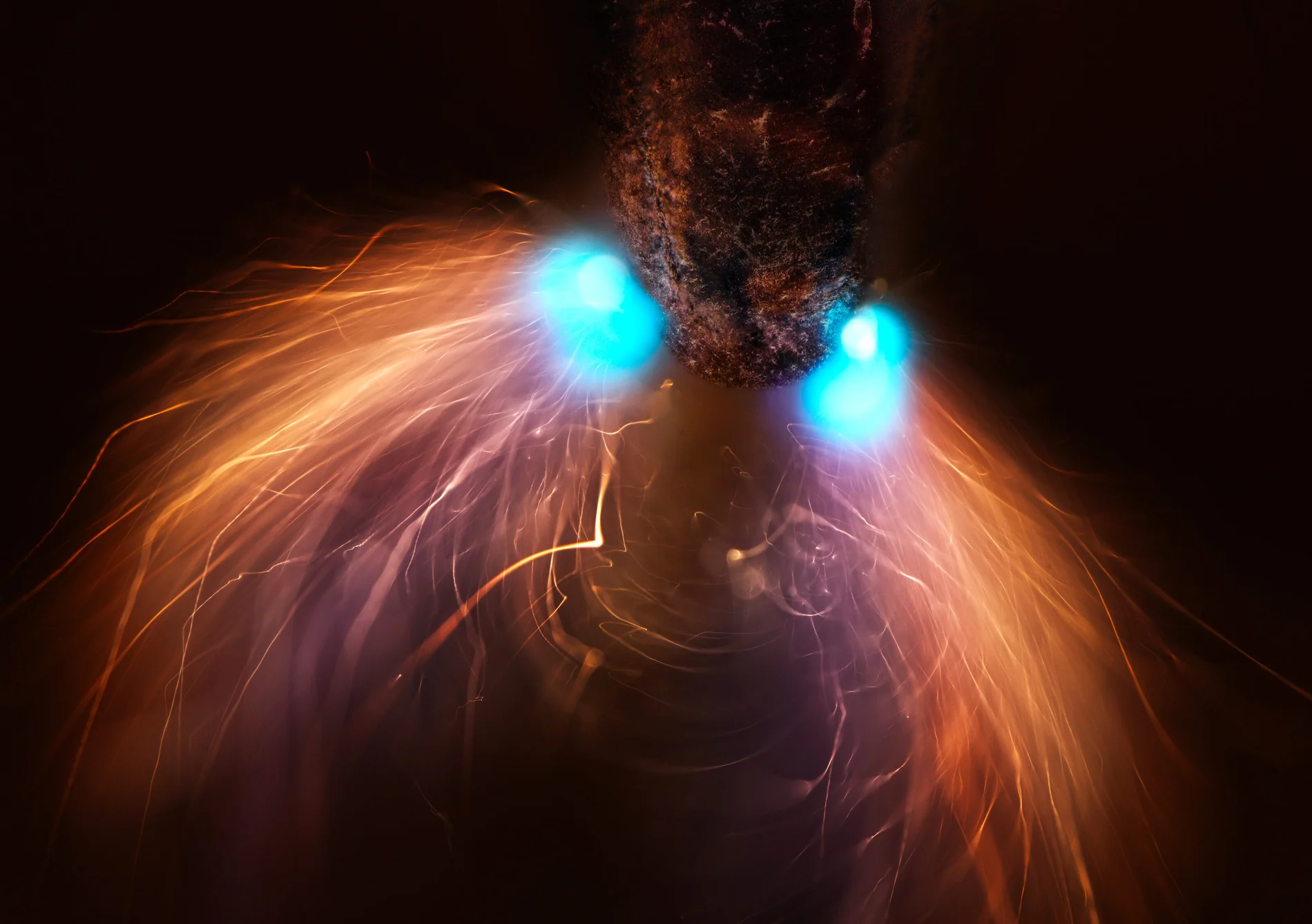
8th place — Marcel Clemens. Pencil lead during high-voltage electrical discharge.
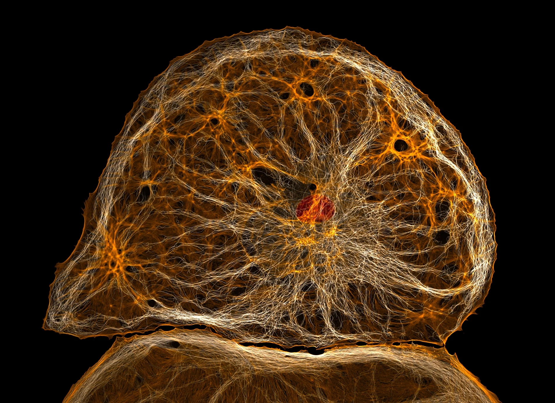
9th place — Francisco Lázaro-Diéguez. Dedifferentiated liver endothelial cell.
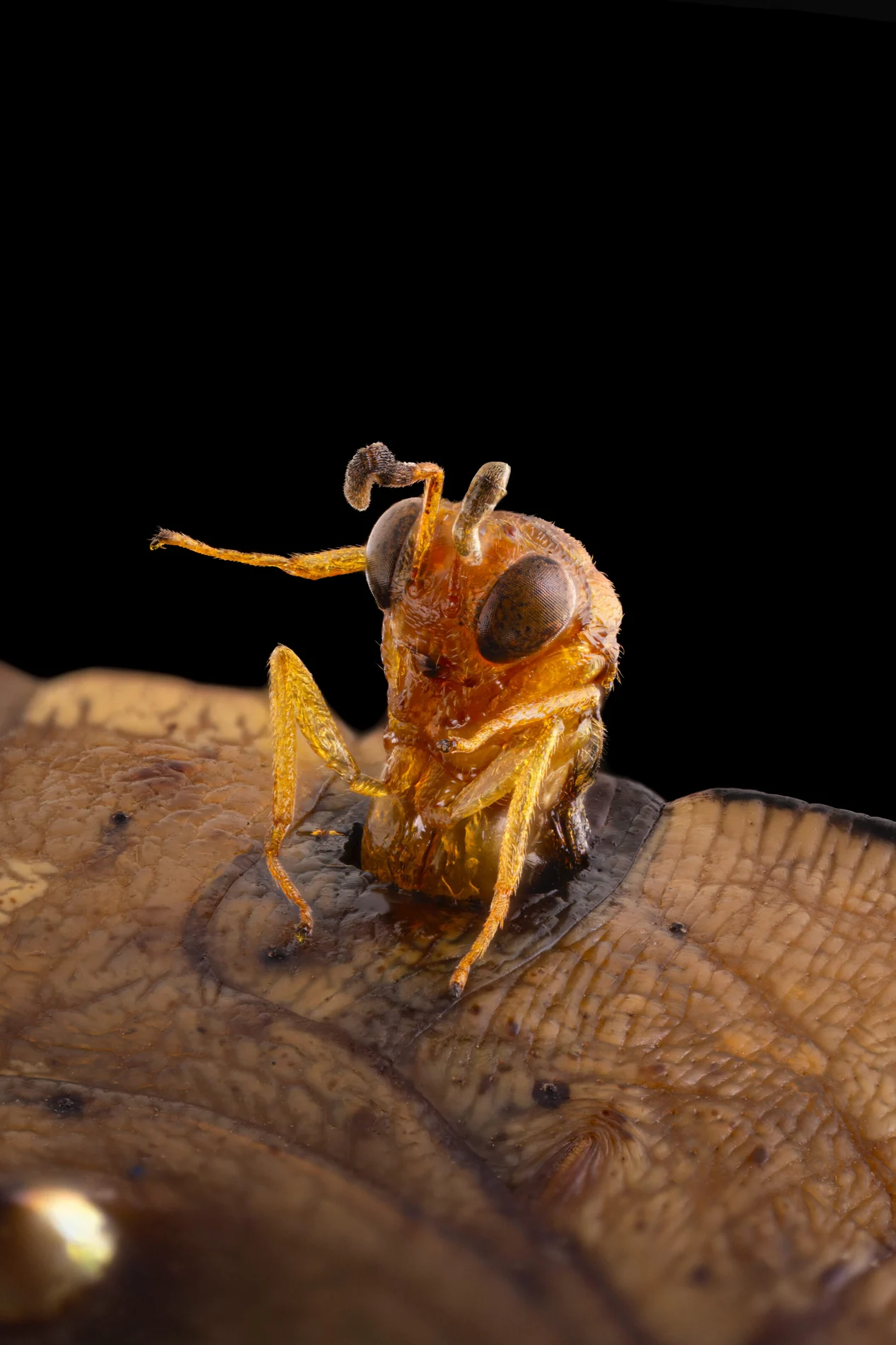
10th place — Michael Parra Puentes. Conura parasitoid wasp emerging from a butterfly chrysalis.
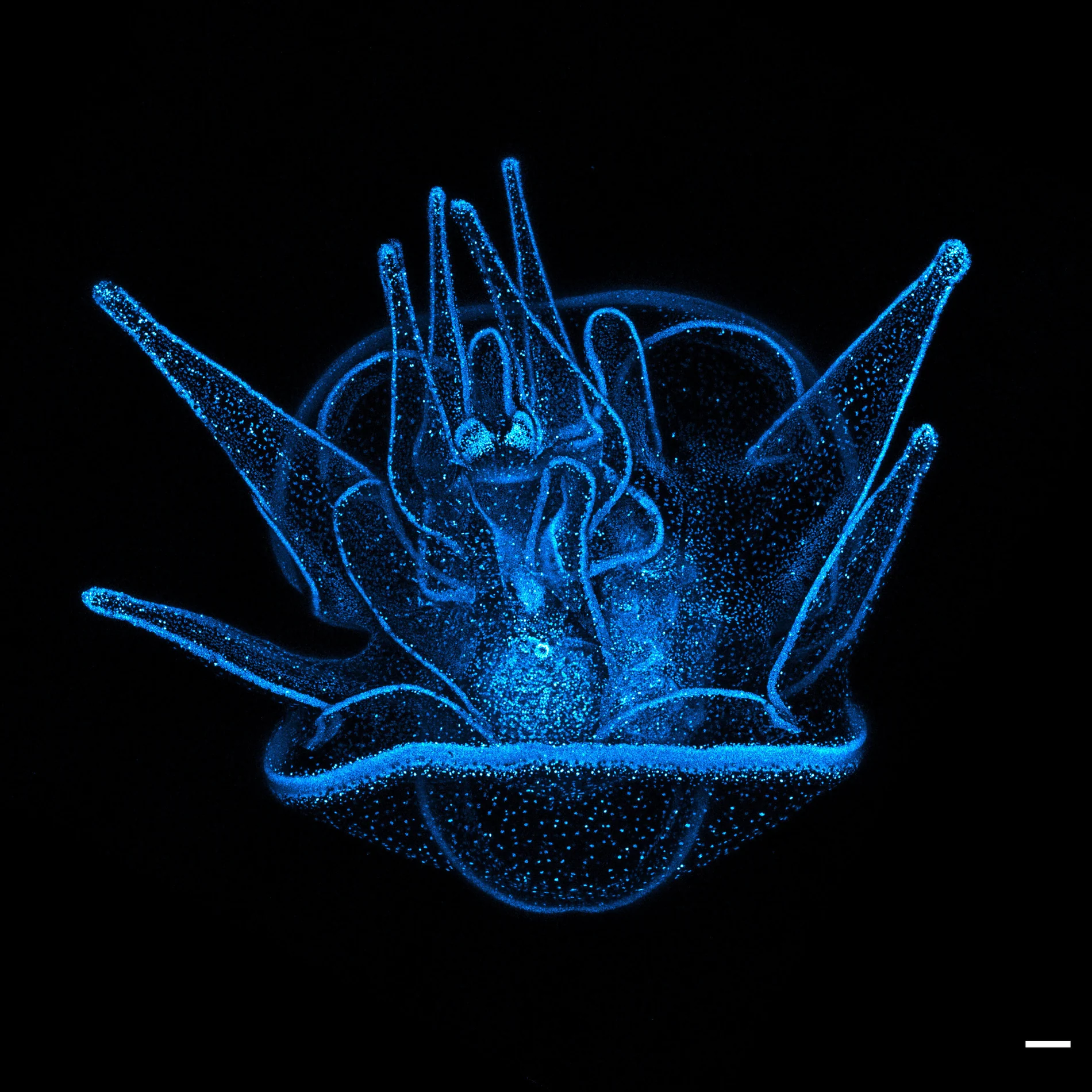
11th place — Svenja Carolin Kling. 20-day-old sea urchin larva with blue fluorescent nuclei.
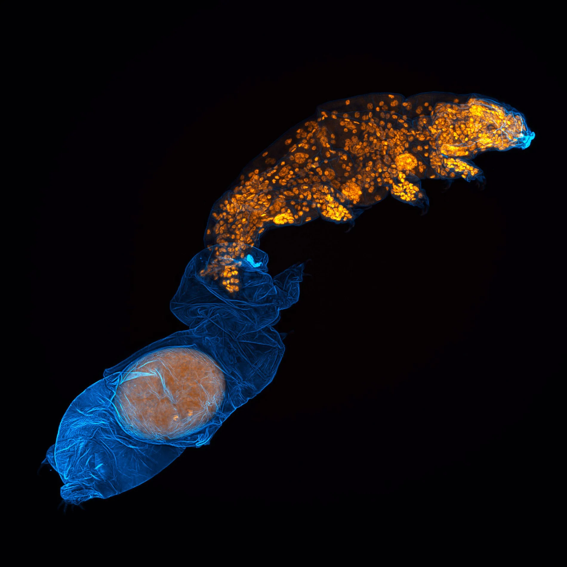
12th place — Gonzalo Quiroga Artigas. Tardigrade molting and laying an egg in its old skin.
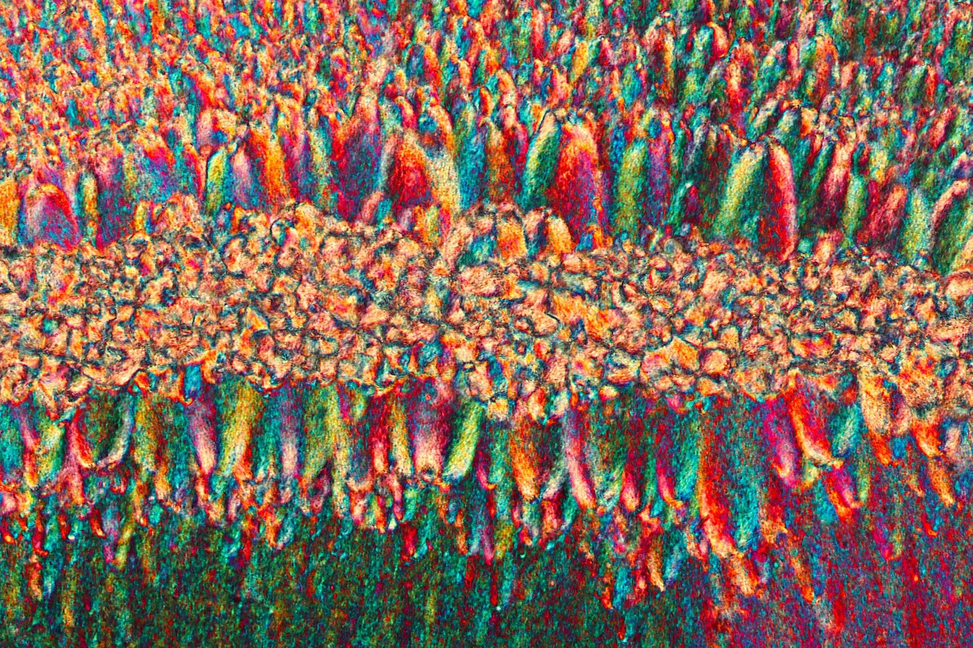
13th place — Sebastian Sparenga. Recrystallized estrogen.
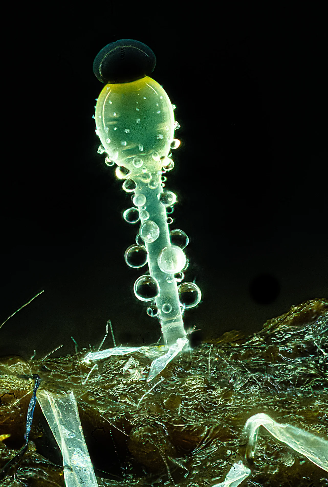
14th place — Mike Crutchley. Pilobolus fungi on sheep dung.
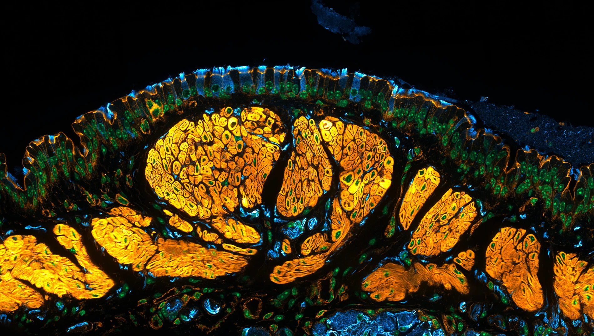
15th place — Julien Resseguier. Mouse trachea with ciliated cells and nerve fibers.
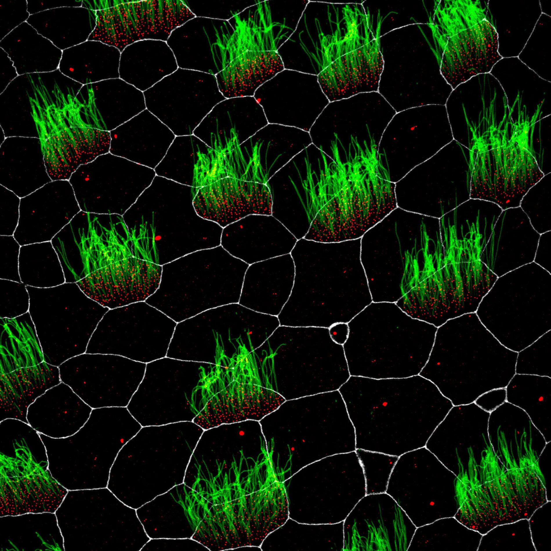
16th place — Pierre Nouaux, Laurent Kodjabachian και Camille Boutin. Embryonic Xenopus skin cells showing cilia, centrioles and membranes.
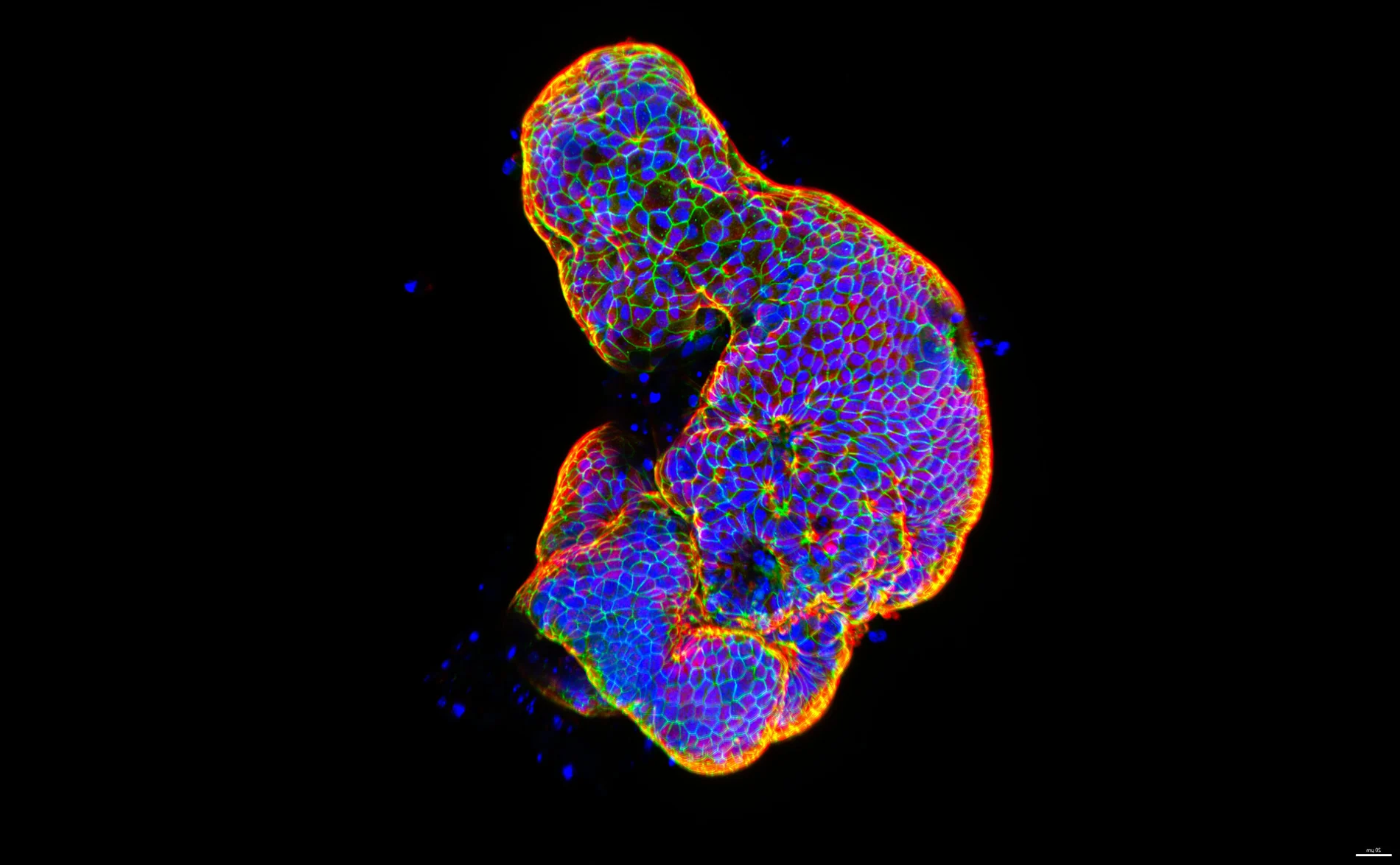
17th place — Gauri Kulkarni. Lab-grown human intestinal tissue.
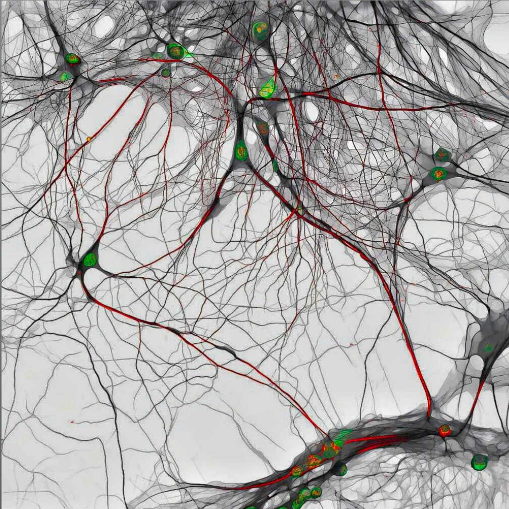
18th place — Bilal Akhtar. 14-day-old mouse hippocampal neurons.
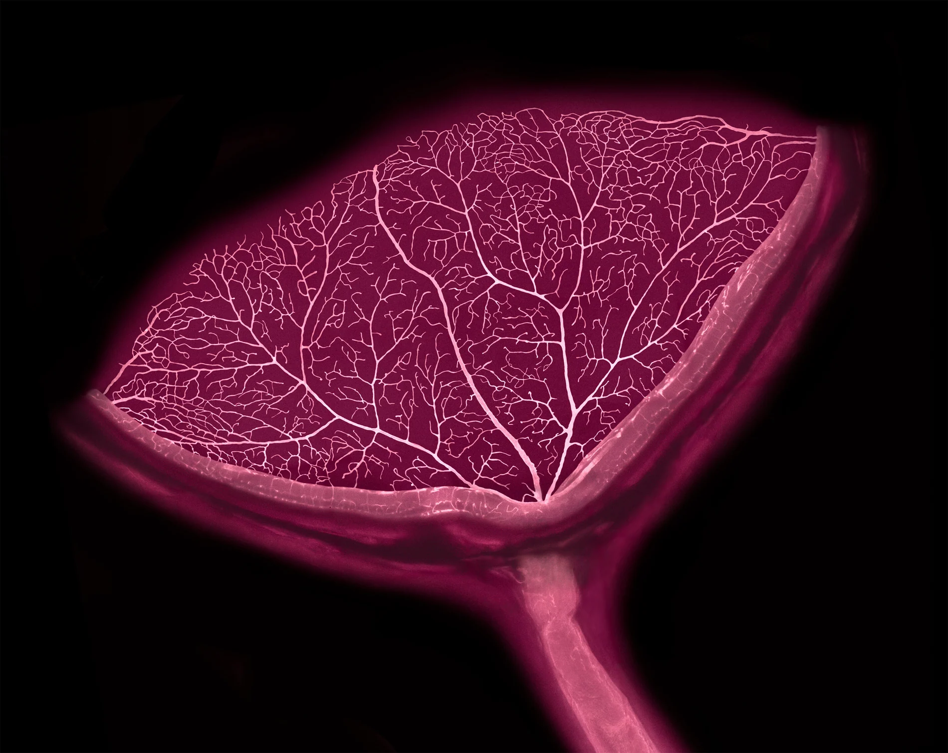
19th place — Satu Paavonsalo και Sinem Karaman. Cross-section of mouse eye with retinal blood vessels and optic nerve.
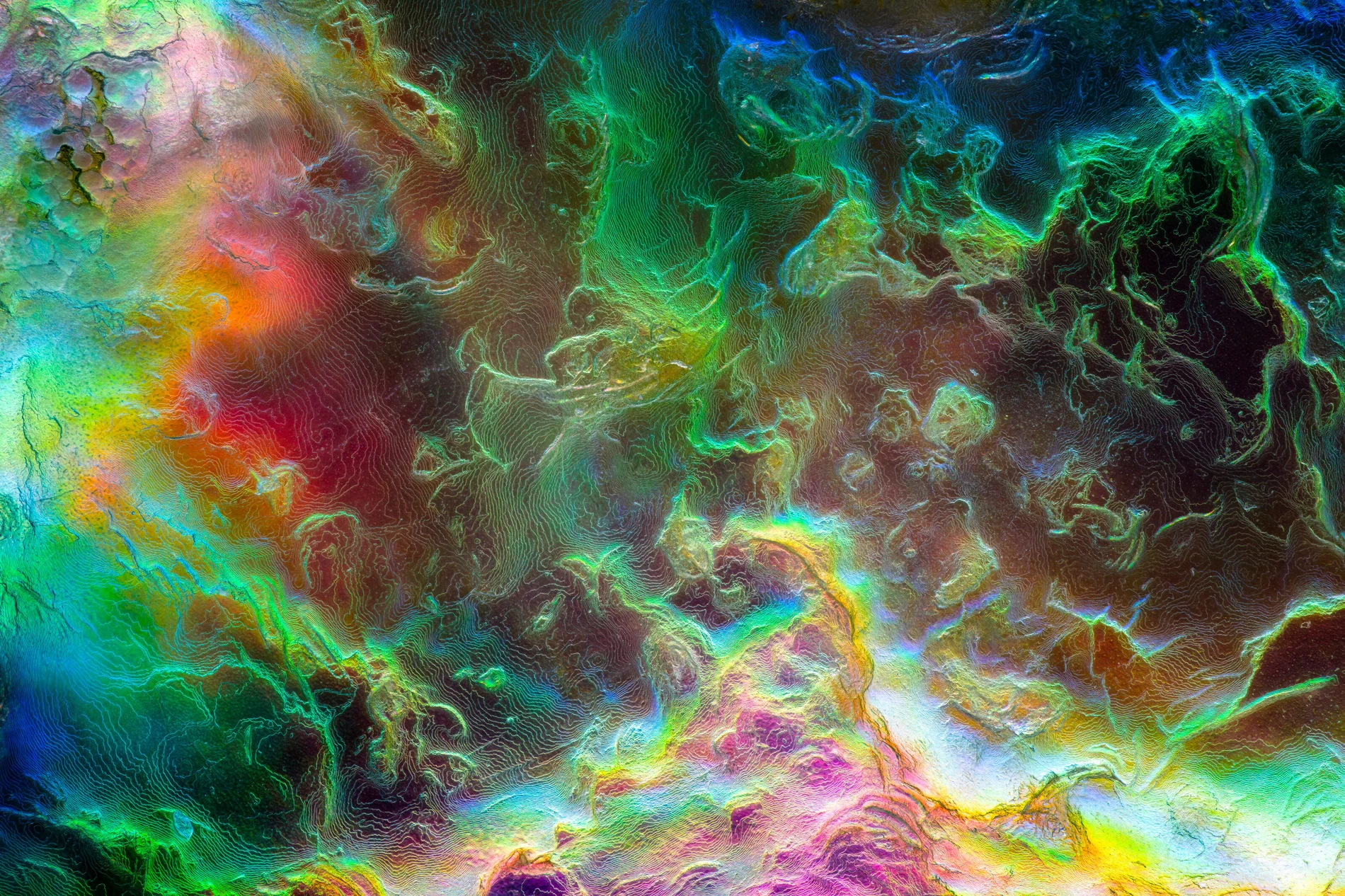
20th place — Justin Theodore Benttinen. Abalone shell.
Nine honorable mentions
Claire J Ang. Mouse uterus with epithelial gland structures.
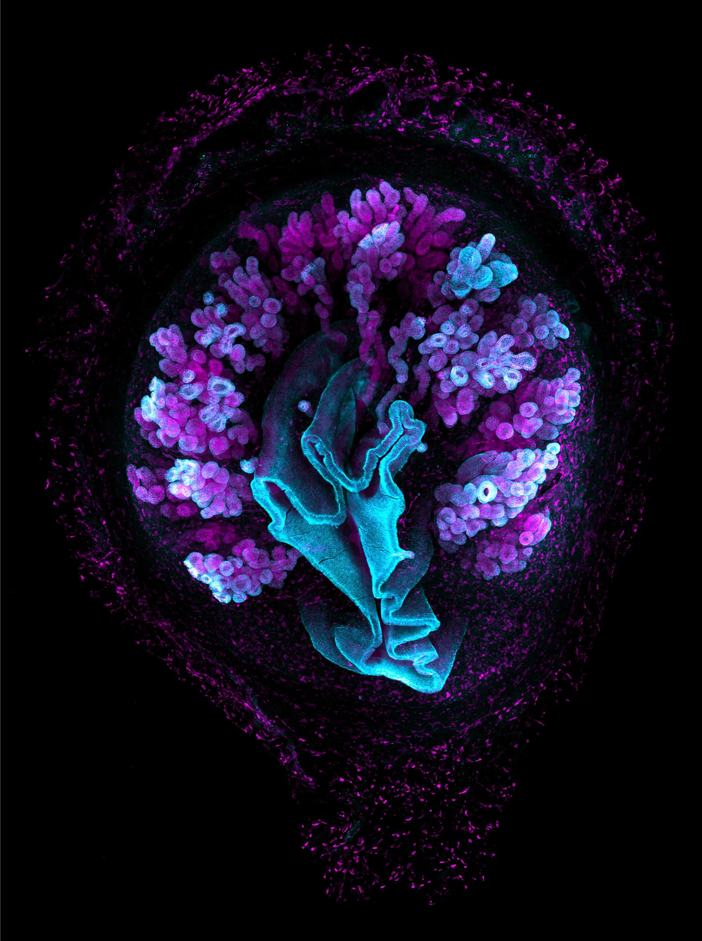
Richard Atherton. Trypanosoma cruzi parasites inside human fibroblasts.
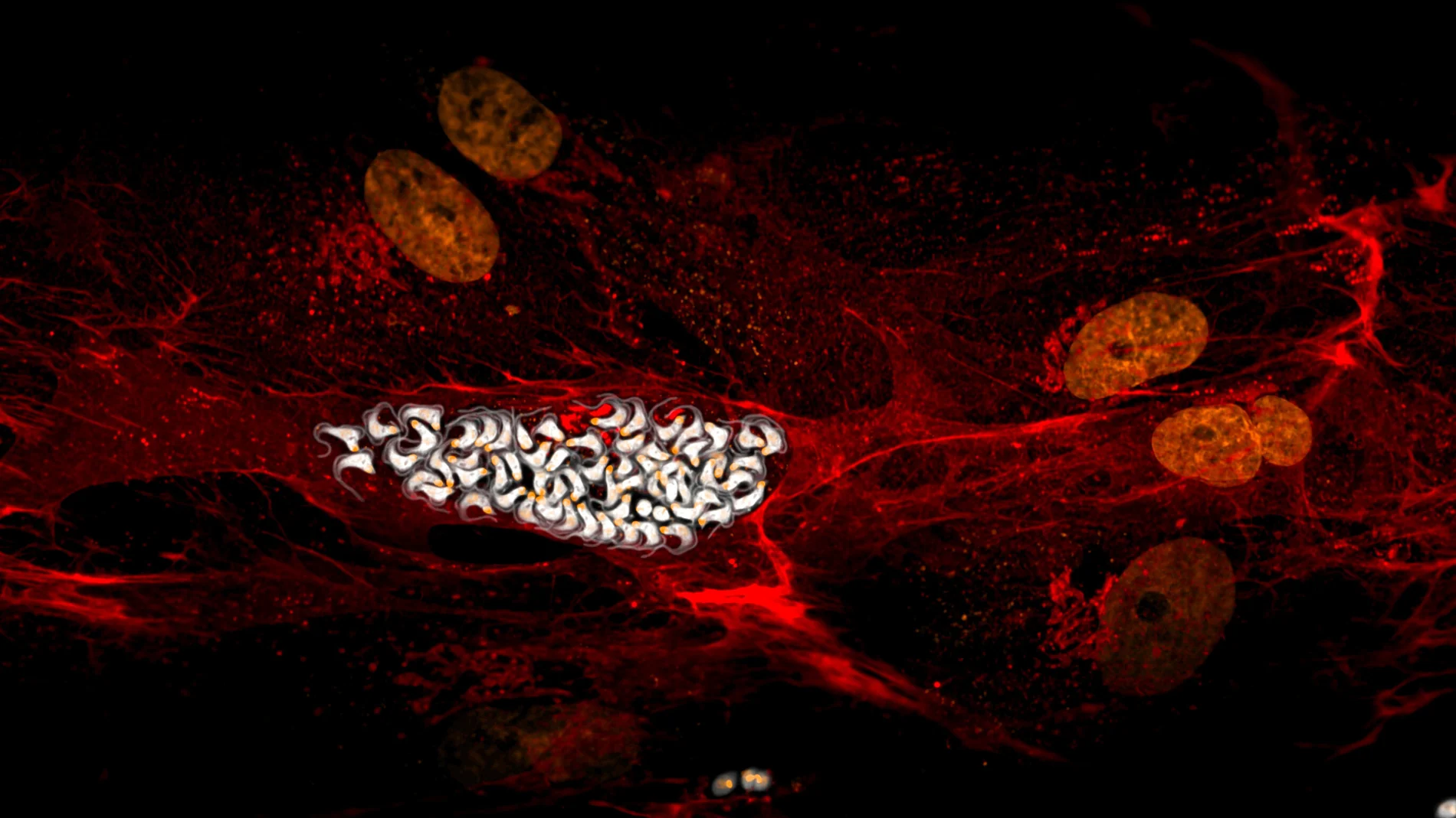
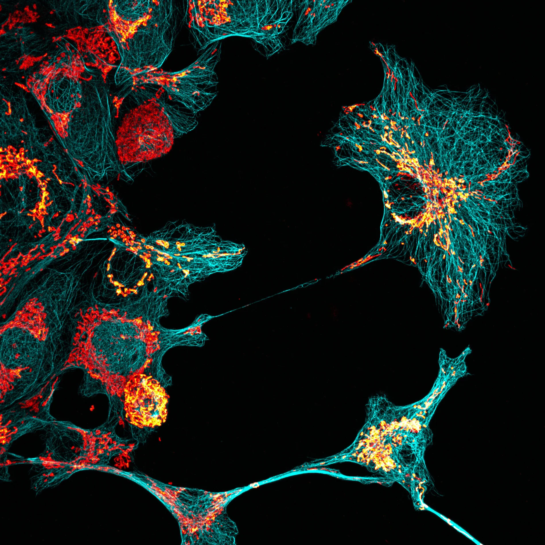
Walid Idi. African green monkey kidney cells with microtubules and mitochondria.
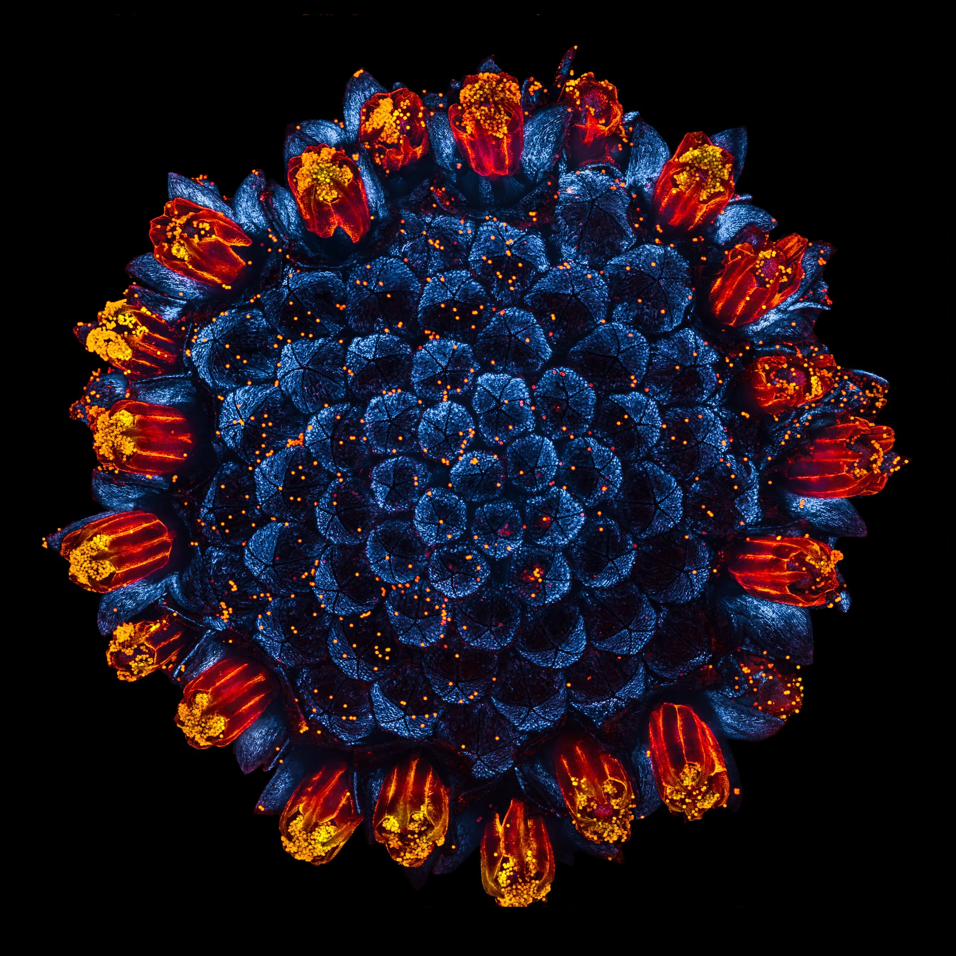
Jan Martinek. Daisy flower head exhibiting natural autofluorescence.
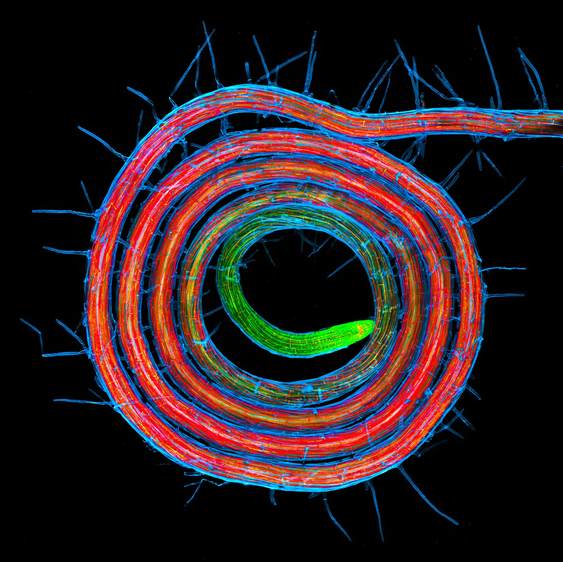
Jan Martinek. Arabidopsis root coil.
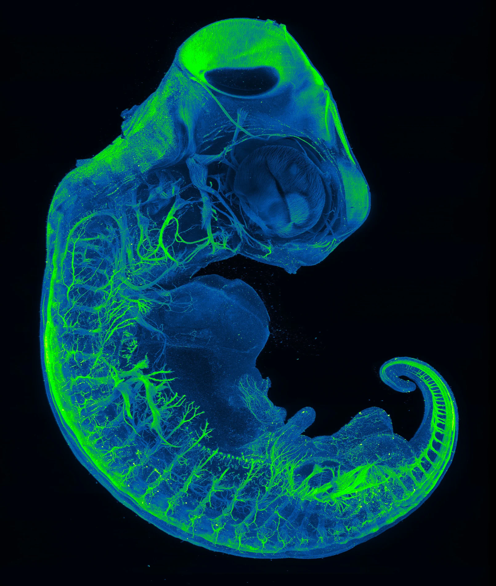
Amartya Tashi Mitra και συνεργάτες. Neurons of a red-eared slider turtle embryo.
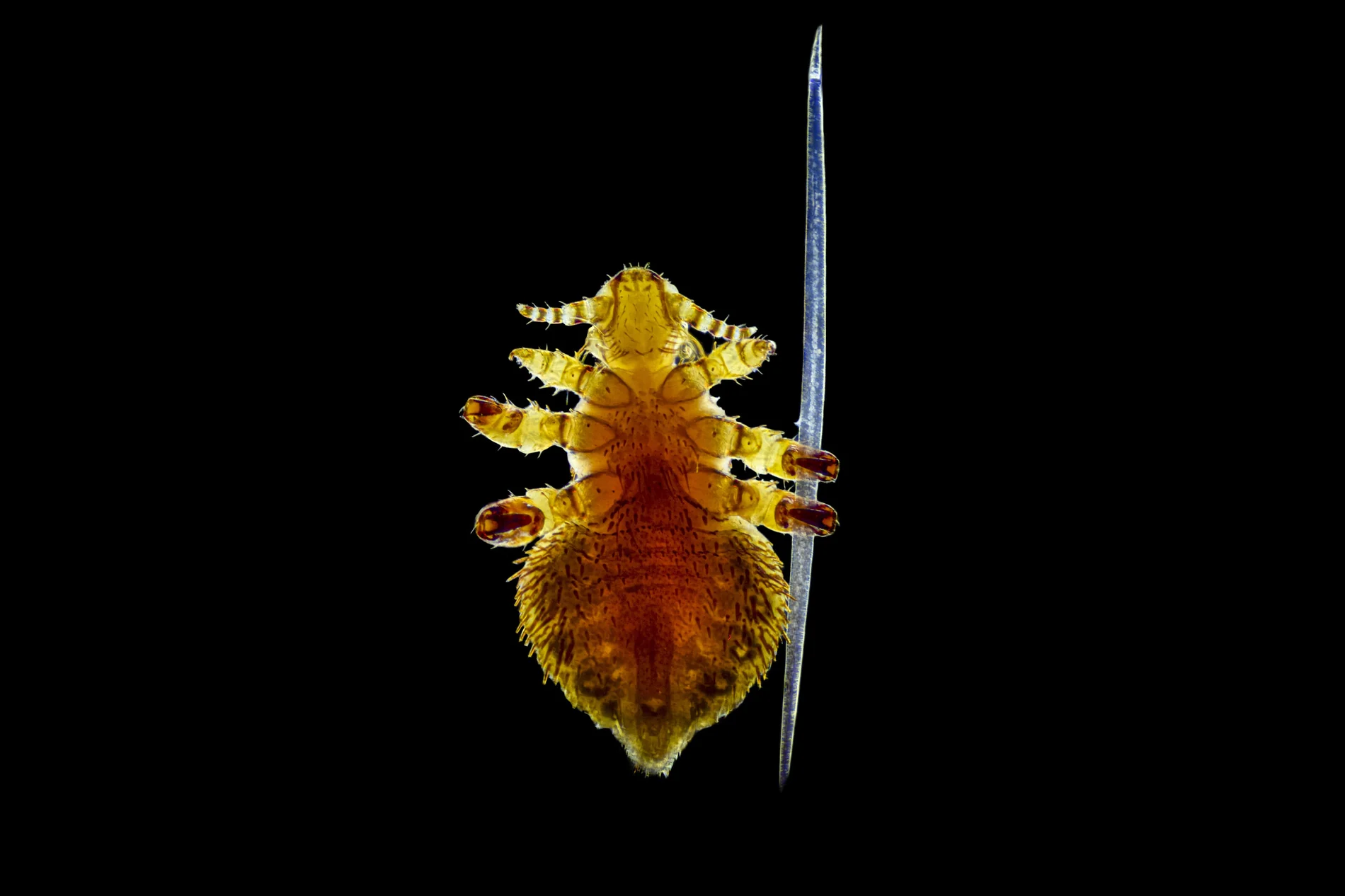
Brandon Antonio Segura Torres και συνεργάτες. Seal louse gripping a single seal hair.
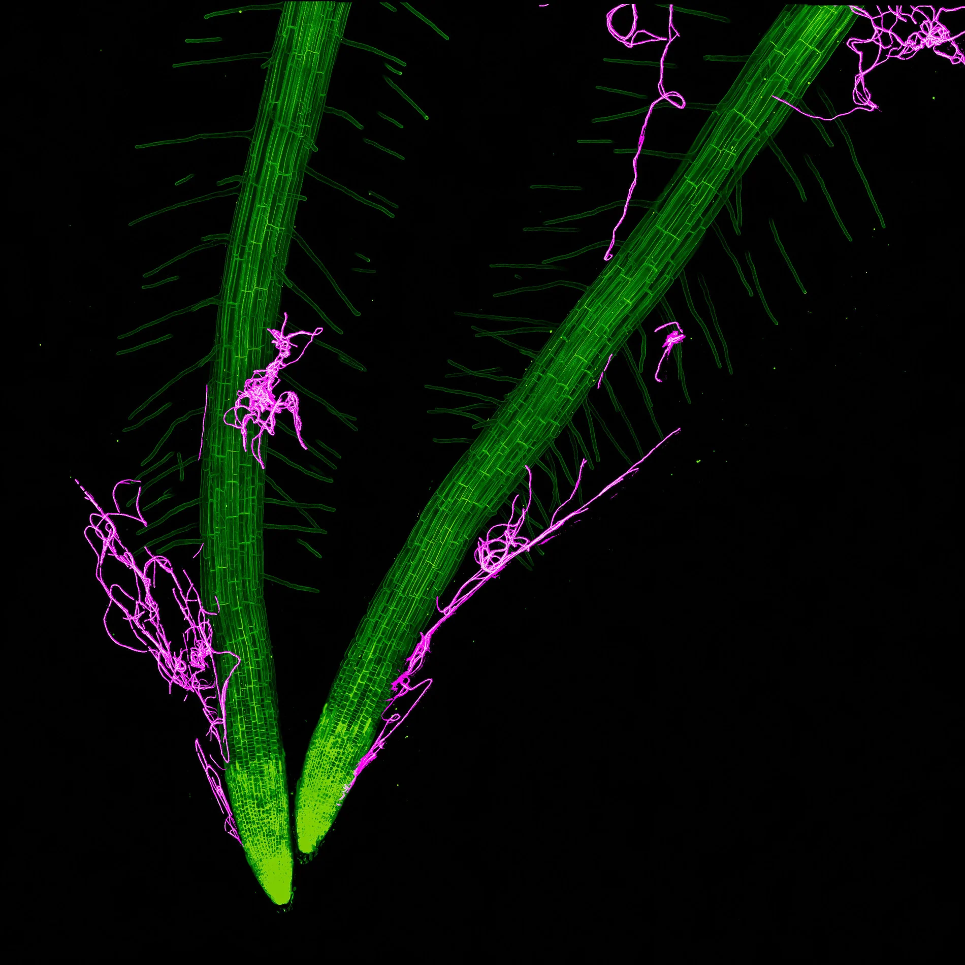
Kevin Y Shih. Arabidopsis roots colonized by Bacillus thuringiensis.
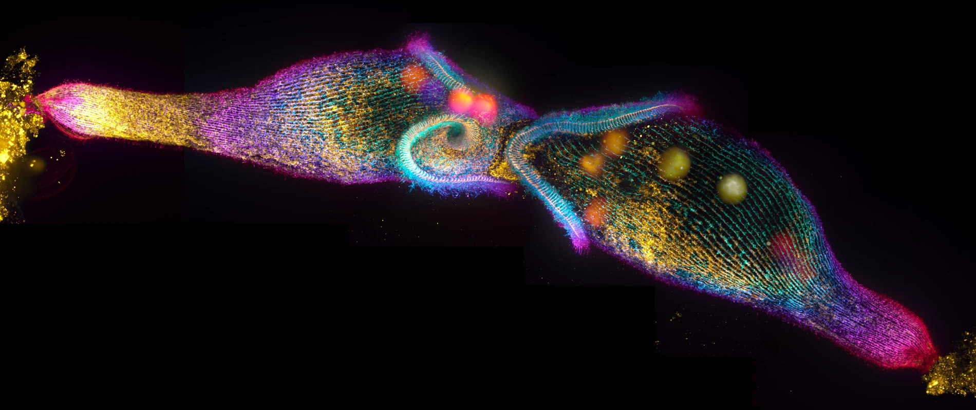
Mark Slabodnick. Two Stentor coeruleus cells mating.
The techniques behind the photographs
The photographs use different methods, including focus stacking, confocal microscopy, fluorescence, darkfield and polarized light. Some combine many focal planes to keep a three-dimensional microscopic subject sharp. Others rely on special markers or properties of light to isolate structures that would otherwise be difficult to distinguish.
The competition has a long history of celebrating microscopic photography. The 2019 Nikon Small World winners already demonstrated how varied the subjects of scientific imaging can be.
What we think
The top photograph is an excellent example of a photographer treating an engineering problem as a creative challenge. Bielfeldt did not need an exotic subject: he chose an ordinary printer, changed the way it worked in front of his lens and found a way to freeze a process that normally happens far too quickly to see.
It is also worth looking at the variety in the remaining photographs rather than thinking of them all as straightforward macro shots. Neurons, cells and crystals are not photographed with identical methods, nor do their colors always represent the same kind of information. For photographers, the magnification, lighting and capture method are part of the story behind the image.
And that is perhaps the most compelling quality of the 2026 selection: alongside sophisticated laboratory methods there is room for a pollen-covered spider, the tip of a pen and a printer firing ink. Curiosity starts the project; good technique makes the image possible.