


Summary
- Google Research introduced PhotoScan, an experimental AI system that estimates body composition from smartphone photos
- The model was initially trained on data from 35,323 UK Biobank participants and fine-tuned on 677 adults
- In independent validation, it achieved a mean absolute error of 2.13 percentage points for body-fat percentage
- Adding PhotoScan data increased AUROC for insulin-resistance classification from 0.692 to 0.760
- DXA data reached an AUROC of 0.773, slightly higher than PhotoScan
- The system remains a research prototype and has not been announced as a consumer or medical feature
Google Research has introduced PhotoScan, an experimental artificial-intelligence system that estimates body composition from smartphone photos and can improve insulin-resistance prediction beyond what Body Mass Index alone provides.
The study shows that information extracted visually from the body can, in a research setting, approach DXA performance for assessing cardiometabolic risk.
Google Research published new results on August 17, 2026, for PhotoScan, a deep-learning system under development that analyzes standard smartphone body photographs and estimates metrics including total body-fat percentage, the ratio of fat stored around the trunk versus the hips, and the ratio of visceral to subcutaneous fat.
The significance of the research is that BMI, although simple and widely used, does not reveal where fat is stored and does not adequately distinguish fat mass from lean mass. PhotoScan attempts to add this information without requiring a specialized scanner, with the aim of making cardiometabolic-risk assessment more accessible.
What PhotoScan actually measures
The system does not simply attempt to estimate weight or BMI from an image. It was trained to predict three more specialized body-composition indicators: body-fat percentage (BF%), the Android-to-Gynoid fat ratio or A/G, and the Visceral-to-Subcutaneous fat ratio or V/S.
The A/G ratio broadly compares fat accumulation around the trunk with that around the hips and thighs. V/S examines the relationship between visceral fat around internal organs and subcutaneous fat under the skin. This distribution can provide more information about metabolic risk than a single overall body-fat percentage.
How the model was trained
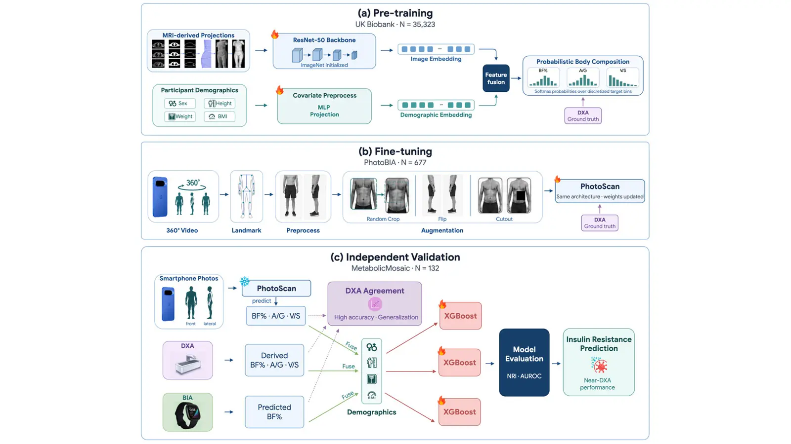
Development took place in three main stages. Google first pretrained a ResNet-50 neural network using data from 35,323 UK Biobank participants. Front and side 2D projections were generated from 3D MRI scans, while DXA measurements were used as body-composition ground truth.
The system was then fine-tuned on real smartphone photographs using the PhotoBIA cohort of 677 adults. The photographs were paired with actual DXA measurements and evaluated with five-fold cross-validation.
Finally, performance was tested on an independent group of 132 people in the MetabolicMosaic cohort. This group participated in a 30-week longitudinal study in San Francisco, with data from DXA, PhotoScan, bioelectrical impedance, anthropometric measurements, fasting blood tests and continuous Fitbit monitoring.
How close it came to DXA
In the PhotoBIA cohort, PhotoScan recorded a mean absolute error of 2.15 percentage points when estimating body-fat percentage. By comparison, the BIA-based model had an error of 2.91.
In the independent MetabolicMosaic cohort, the mean absolute errors were 2.13 for body-fat percentage, 0.085 for A/G and 0.085 for V/S. Researchers described the results as consistent across the fine-tuning and independent-validation datasets.
The comparison matters because Dual-Energy X-Ray Absorptiometry is considered one of the most accurate methods for assessing body composition, but it requires specialized clinical equipment and involves a small dose of radiation.
The biggest difference appeared in insulin resistance
Researchers did not stop at estimating body fat. They used PhotoScan data to examine whether it could help classify people with insulin resistance.
A baseline model using age, sex and BMI achieved an AUROC of 0.692. Adding PhotoScan body-composition metrics increased that figure to 0.760, with an NRI of 0.593. According to the paper, the AUROC improvement was statistically significant.
Using DXA data produced an AUROC of 0.773 and an NRI of 0.748. In other words, PhotoScan approached the performance of clinical DXA in this particular research test, although it did not surpass it.
By contrast, Google says adding smartwatch BIA measurements did not improve AUROC or NRI over the baseline demographic model in the same evaluation. The researchers attribute part of the difference to the fact that BIA mainly provides an estimate of total body-fat percentage, while PhotoScan also attempts to estimate fat distribution.
Why BMI is not always enough
BMI is calculated from weight and height and remains a useful population-level indicator, but it does not directly describe body composition.
Two people with similar BMI values can have different muscle mass, different body-fat percentages and, importantly, different fat distribution. Google’s research is based on the idea that the additional geometric information visible to the camera can provide clinically useful signals that are missed when only BMI is considered.
It is not an app consumers can use today
PhotoScan remains a research prototype. Google itself describes it as an investigational framework and has not announced commercial availability or integration into Pixel, Fitbit or another health service.
The results also do not mean that a simple photograph can replace clinical diagnosis or laboratory testing. This work evaluates the ability of a model to estimate body-composition indicators and contribute to risk-classification models in controlled research datasets.
Google also stresses that body composition is only one component of cardiometabolic health and says future work will explore combining it with wearable data, glucose dynamics and clinical blood biomarkers.
Smartphone photos are becoming biometric sensors
The interesting part is not only the accuracy of this particular model, but the broader shift in the role of the camera. A smartphone camera is gradually moving beyond being purely an image-capture device and can serve as a sensor from which machine-learning systems extract information that is not immediately visible to the user.
Google has already invested heavily in AI-based visual understanding. PTTL has previously covered Google’s ability to answer questions about what a smartphone camera sees in real time, a different application of the same broader direction: the image becomes a source of data rather than simply a photograph.
In health applications, however, that transition brings much higher requirements for reliability, privacy and clinical validation because body images and the conclusions that can be drawn from them represent highly sensitive information.
What we think
PhotoScan is one of the more interesting indications of how computational photography could move beyond improving an image and become a measurement tool.
The results are noteworthy, particularly because the system approached DXA performance in a specific insulin-resistance classification test. Research performance should not, however, be confused with a finished medical product. PhotoScan’s real value will depend on much broader independent validation, its performance across different populations and shooting conditions and, if it ever reaches consumers, how privacy and the handling of highly sensitive visual data are addressed.
Frequently asked questions
What is the Google PhotoScan in this research?
It is an experimental deep-learning system from Google Research that estimates body-composition metrics from smartphone photos. It should not be confused with older Google products carrying a similar name.
Can it replace a DXA scan?
No. The research shows that PhotoScan approached DXA-derived performance in a specific insulin-resistance prediction test, but DXA remains the more accurate clinical method and PhotoScan is still a research prototype.
Is it available on Pixel or other smartphones?
No. As of August 20, 2026, Google has not announced a commercial PhotoScan feature for Pixel, Fitbit or another consumer service.
How accurate was body-fat estimation?
In the independent MetabolicMosaic cohort, the mean absolute error for body-fat percentage was 2.13 percentage points. In the PhotoBIA cohort it was 2.15.
Why does fat distribution matter more than BMI alone?
BMI does not reveal where fat is stored and does not accurately distinguish fat mass from lean mass. Measures such as A/G and V/S can provide additional information about fat distribution and cardiometabolic risk.