

Summary
- Canon and Coral Spawning International used medical ultrasound to detect developing eggs inside living corals.
- The Aplio i700 was used on six hard-coral species.
- Researchers compared ultrasound images with macro photography to validate their findings.
- The team will monitor the corals for six months to better understand their spawning cycles.
A Canon medical ultrasound scanner has been used to find developing eggs inside living corals, giving scientists a way to follow their reproduction without cutting the animals open.
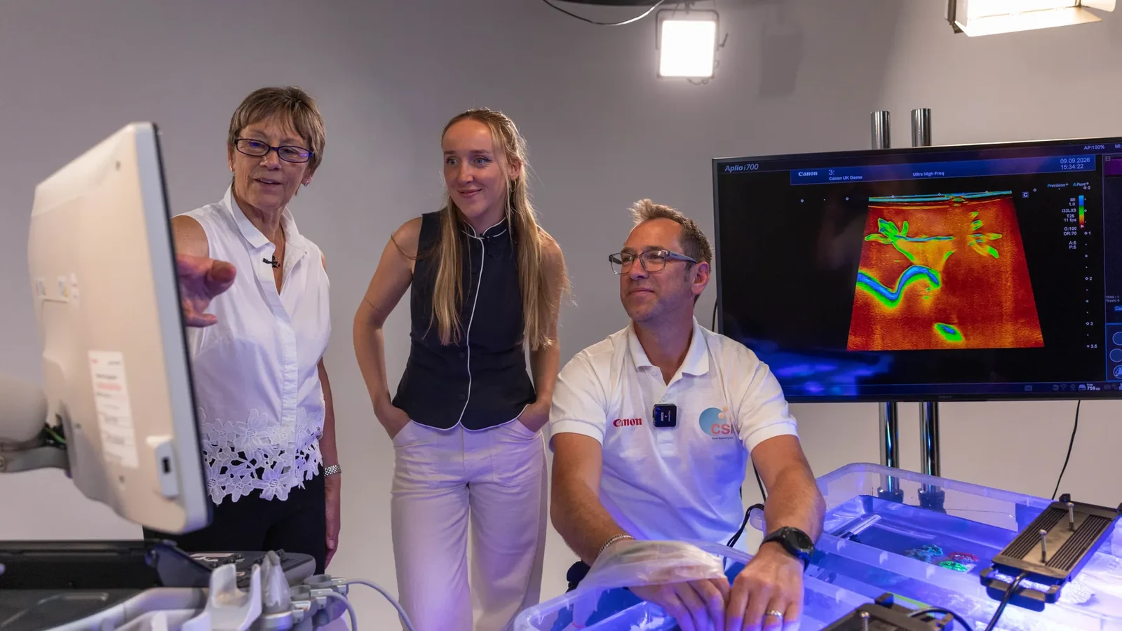
Coral Spawning International (CSI), Canon Medical Systems UK and Canon EMEA announced on November 4, 2026, what Canon describes as the first such ultrasound examination of living, submerged corals. The experiment forms part of the company’s World Unseen initiative.
Taking a hospital ultrasound scanner into a coral tank
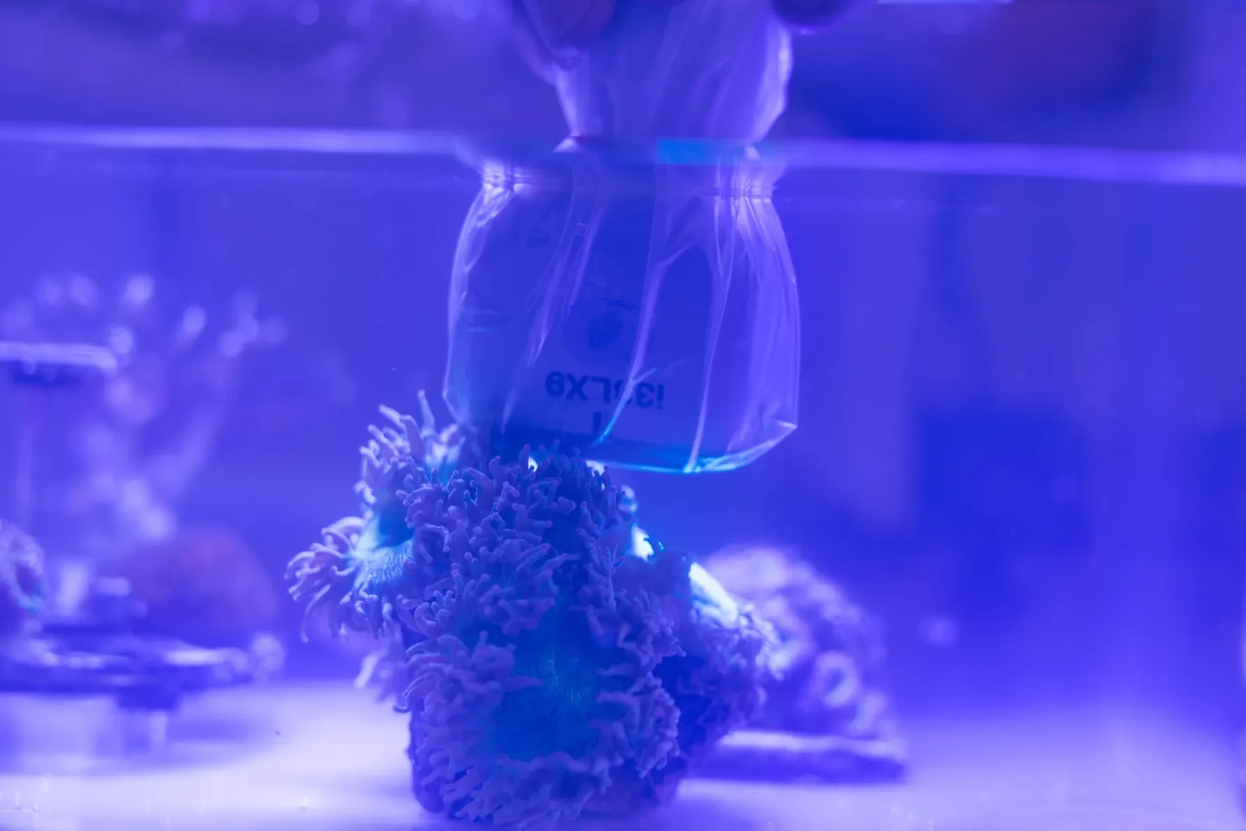
The team used the Canon Aplio i700, a system normally found in hospitals for imaging human tissues and organs. This time, they scanned six species of large-polyp stony (LPS) coral with different skeletal structures, keeping the corals fully submerged throughout the examination.
To resolve their fine internal structures, the researchers combined high-frequency and ultra-high-frequency probes. The former offer deeper penetration, while the latter reveal finer details closer to the probe. That balance matters when the target is a small cluster of eggs behind different layers of tissue and skeleton.
More information here about Canon Medical Systems’ Aplio i700.
What the ultrasound and macro photograph reveal
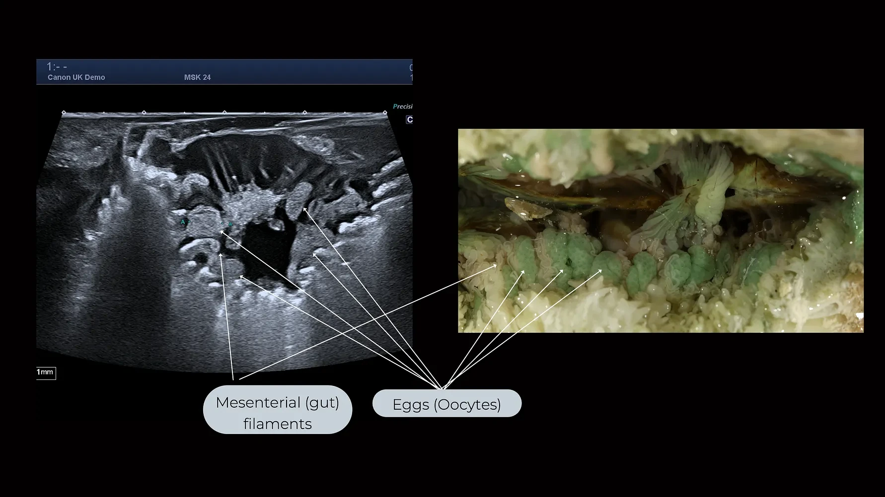
The key result was the detection of developing eggs inside living coral polyps. In the comparison image, the scientists placed the ultrasound scan alongside a macro photograph showing the internal anatomy of a Trachyphyllia geoffroyi coral. Arrows identify the oocytes and nearby mesenterial filaments.
The comparison helped verify what the ultrasound was actually showing. Direct examination of internal tissue was also involved at this validation stage; the advance is that subsequent ultrasound observations can be performed without destructive sampling. That makes repeat measurements on the same living corals possible.
Why the timing of coral reproduction matters
Corals are animals, and they release reproductive cells during particular spawning windows that vary with the species and environmental conditions. Knowing when eggs are maturing can help researchers time gamete collection and controlled breeding activities used in reef restoration programmes.
This behaviour is so difficult to capture that a rare photograph of a coral spawning stood out among the Underwater Photographer of the Year 2026 category winners. Ultrasound adds something different to visual documentation: information about what is happening before the visible release of reproductive cells.
The team plans to keep monitoring the corals for six months, including recording the release of gametes. This should help them assess how useful ultrasound imaging is for predicting spawning windows across different coral species.
What we think
What makes this particularly compelling is that imaging here does more than produce a striking picture. It gives marine biologists information that was difficult to obtain without interfering with the animal itself. Being able to return to the same coral means they can watch a process unfold rather than capture just one isolated moment.
For anyone involved in nature photography, the side-by-side images make an interesting comparison. Macro photography reveals extraordinary detail on a surface or an exposed specimen, but ultrasound shows what is happening inside a living animal. These are not competing forms of imaging: one helps scientists understand what they are seeing in the other.
Protecting coral reefs takes much more than a new imaging technique. Still, knowing when a coral is preparing to reproduce without destroying it in the process is a concrete and useful capability for the researchers trying to keep these ecosystems alive.
Frequently asked questions
Are corals plants?
No. Corals are animals made up of polyps. The researchers used ultrasound to examine reproductive structures inside living coral polyps.
Does the ultrasound examination damage the coral?
Ultrasound scanning does not require the coral to be cut open. The initial validation involved comparing the scan with direct examination of internal tissue, but the purpose of the technique is to allow subsequent non-invasive examinations.